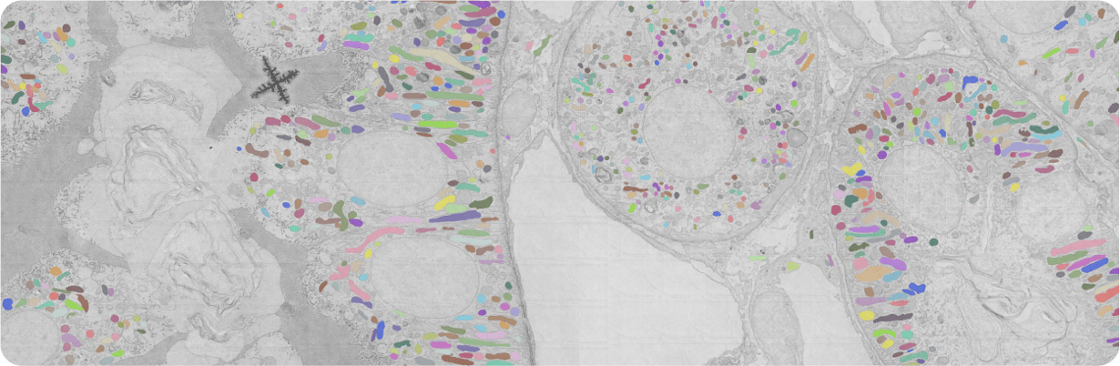
Discover how an artificial intelligence (AI) based mitochondrial model together with the FAST-EM can be used to automatically identify mitochondria in hundreds of cells, allowing for morphometrics extraction.
The dysregulation of mitochondria, dynamic organelles essential for cellular health, are associated with various diseases. Electron microscopy (EM) can reveal morphological details of mitochondria, offering insights into state of the cell population health. While conventional EM is relatively slow and has to compromise between resolution and image area, Delmic’s FAST-EM overcomes this, capturing high-resolution images of thousands of mitochondria within minutes rather than hours. Download our whitepaper to learn about an easy-to-use automated segmentation model for FAST-EM data, enabling quantitative and automated analysis of mitochondria in hundreds of cells.
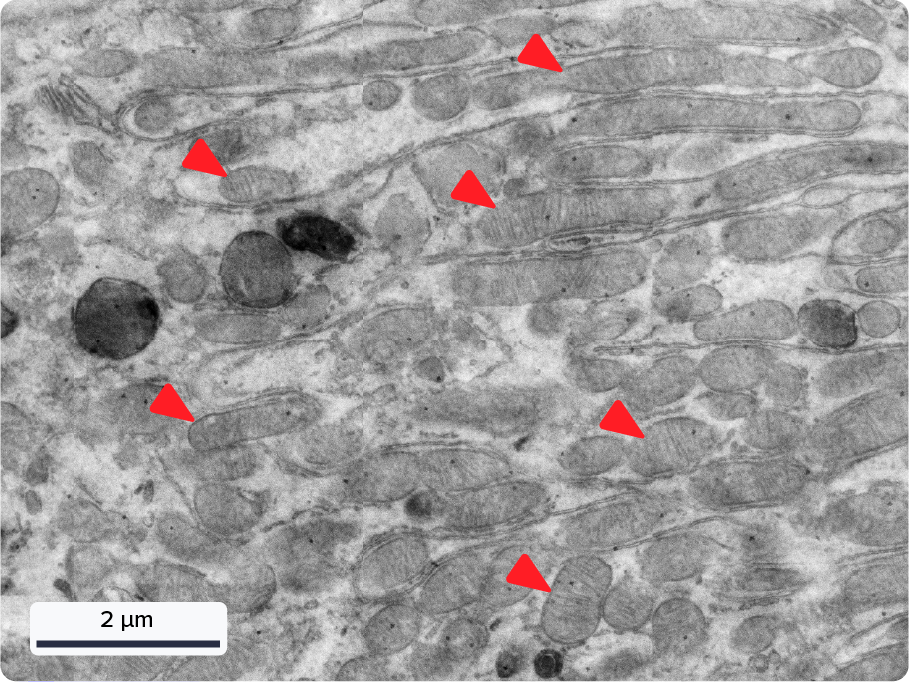
Figure: A zoomed-in view of a FAST-EM image of a healthy rat kidney sample. At a 4 nm pixel size, the resolution is sufficiently high to visualize the cristae in individual mitochondria (red arrows).
.svg "DELMIC")